Recently, research group led by Professor Zhong Guisheng at iHuman institute and School of Life Science and Technology, with colaberators Professor He Shuijin at School of Life Science and Technology, Professor Chai Renjie at Southeast University identified novel cytoskeleton ultrastructures in inner ear hair cell utilizing super-resolution fluorescence microscope. Their two studies, “Critical role of spectrin in hearing development and deafness”, and “A cytoskeleton structure revealed by super-resolution fluorescence imaging in inner ear hair cells” have been published in Science Advances (Beijing time: Apr 18, 2019, on-line published) and Cell Discovery (Beijing time: Feb 19, 2019) successively.
Hearing loss is the most common sensory disorder world-wild. Over 6.8% of the global population (around 500 million people) suffer from disabling hearing loss and no efficient treatment is yet available. Hair cell dysfunction is one of the most common genetic causes of hearing loss.
The cuticular plate of hair cells (HCs) is thought to be critical in mammalian hearing by securing stereocilia rootlets in place and providing them with the rigidity and support necessary for auditory transduction. Stereocilia rootlets are electron-dense structures penetrating into the cuticular plate and forming an anchoring complex. This anchoring complex is believed to be the key structural component for stereocilia to withstand constant mechanical stresses, and thus plays a critical role in hearing. However, the specific anchoring molecules that provide stereocilia rootlets with their necessary elasticity and flexibility remain elusive.
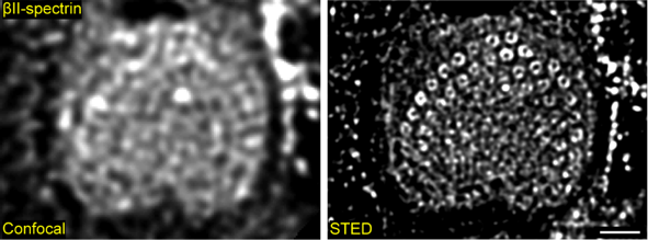
Utilizing stimulated emission depletion (STED) imaging and multiple approaches, a novel ring-like structures of spectrin have been revealed for the first time, wrapping around the base of stereocilia rootlets. These spectrin rings were closely associated with the hearing ability of mice. HC-specific, βII-spectrin knockout mice displayed profound deafness.

Fig 1. STED reveals ultrastructures of βII-spectrin in inner hair cells

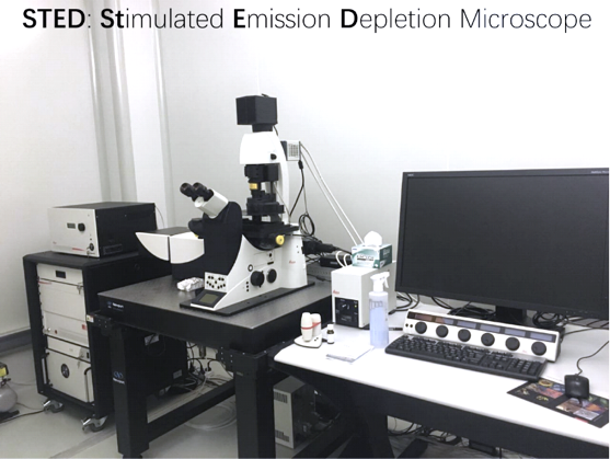
Fig 2. Super-resolution facilities at imaging platform of iHuman institute

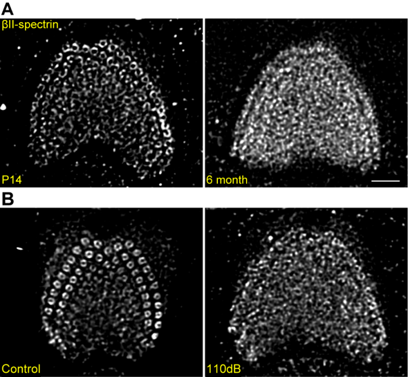
Fig 3. βII-spectrin rings disrupted in the hair cells of aging mice and noise-exposed mice
Further studies have characterized a previously unknown F-actin nanoscale structure in the cuticular plate with structured illumination microscopy (SIM) imaging. Such spatial organization of F-actin in cuticular plate may also play a critical role in hearing function.

Fig 4. Structure of F-actin in the curticular plate of inner ear hair cells
Dr. Zhong Guisheng and his research team have long been committed to studies in the nanostructure of cytoskeleton proteins. The current work for the first time revealed ultrastructures of critical cytoskeletons in inner ear hair cells, and would greatly enhance our understanding of the ultra-structure of cuticular plate and its essential roles in the hearing development.

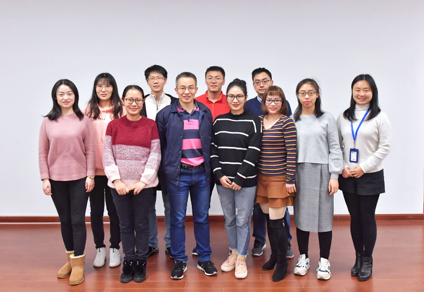
Fig. 5 Research team members of iHuman institute, ShanghaiTech University
Key co-authors of these papers include research assistant professor Liu Yan, doctoral students Qi Jieyu and Chen Xin. Professor Zhong Guisheng, Professor Chai Renjie and Professor He Shuijin are co-corresponding authors.
Cover illustrated by Julie Liu
Read more at:
https://advances.sciencemag.org/content/5/4/eaav7803.full
https://www.nature.com/articles/s41421-018-0076-4

